|

WATSON, J. D. & CRICK, F. H. C.,
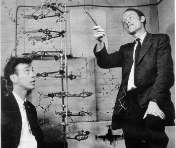
Foto by Antony Barrington Brown 1953
Medical Research Council Unit for the Study of Molecular Structure
of Biological Systems, Cavendish Laboratory, Cambridge.
The complete electronic version of this article can be found online
at
http://www.nature.com/genomics/human/watson-crick/
A Structure for Deoxyribose Nucleic Acid
We wish to suggest a structure for the salt of deoxyribose nucleic
acid (D.N.A.).
This structure has novel features which are of considerable biological
interest.
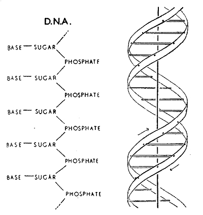
This figure is purely diagrammatic.
The two ribbons symbolize the two phophate-sugar chains,
and the horizonal rods the pairs of bases holding the chains together.
It has not escaped our notice that the specific pairing we have
postulated immediately suggests
a possible copying mechanism for the genetic material.
GENETICAL IMPLICATIONS OF THE STRUCTURE OF DEOXYRIBONUCLEIC
ACID
Nature
171, 964-967 (1953)
By J. D. WATSON and F. H. Crick
Medical Research Council Unit for the Study of the Molecular Structure
of Biological Systems, Cavendish Laboratory, Cambridge

Fig.1 Chemical formula of a single Fig.2
This figure is purely diagrammatic.
chain of Deoxyribonucleic
acid The
two ribbons symbolize the twophosphatesugar chains,
and the horizontal rods the pairs of bases
holding the chains together.
The
vertical line marks the fibre axis.
 |
 |
|
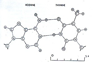
Fig.4 Pairing of adenine and thymine.
Hydrogen bonds
are shown dottet.
One carbon
atom of each sugar is shown
|
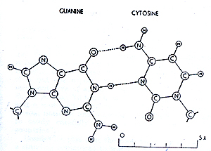
Fig.5 Pairing
of guanine and cytosine.
Hydrogen
bonds are shown dottet.
One carbon atom of each sugar is shown)
|
For the moment, the general sheme we have proposed for the reproduction
of the deoxyribonucleic acid must regard speculative.
Despite these uncertainties we feel that our proposed structure
for deoxyribonucleic acid may help to solve
one of the fundamental biological problems - the molecular basis
of the template needed for genetic replication.
|


