|  
             Institute for Immunology and Genetics. Ger man 
              Cancer Research Centre. Im Neuenheimer Feld 280, 6900 Heidelberg, 
              FRG Henry Kaplan Award for the best poster Immunological Session 
              
              Introduction 
             The thymus represents a crucial phase in the differentiation of 
              T cells, from their earliest precursor committed to the T-cell lineage 
              in the bone marrow, to the full array of peripheral T-effector cells. 
              Specification of T-cell subsets, generation of receptor diversity. 
              selection of self-MHC-restricted T -cell precursors, and induction 
              of selftolerance are thought to be largely or exclusively intrathymic 
              events [24]. How such complex functional events relate to the relatively 
              simple structure of the thymus is poorly understood. It has become 
              apparent that the pattern ofT-cell reactivity is selected by the 
              environment [6, 26] in which T cells develop rather than being strictly 
              genetically fixed; thus interest has focused on the definition of 
              such selection sites. To this end direct cell-cell interactions 
              between cells of the T -cell lineage and nonlymphoid stromal cells 
              in the murine thymus have been characterized [25, 13. 14]. These 
              interactions preexist in vivo, can be isolated as intact multicellular 
              lymphostromal complexes by differential digestion of the thymus. 
              and are thus amenable to analysis in vitro. At least three lymphostromal-cell 
              interactions can be discerned: ( I) between T cells and macrophages 
              (MØ), (2) between T cells and dendritic cells (DC) (both 
              I and 2 referred to as thymocyte rosettes, T-ROS). and (3) between 
              T cells and epithelial cells (thymic nurse cells, TNC). T -ROS and 
              TNC are obtained as sequential fractions during digestion, thereby 
              making possible a separate isolation of distinct complex types. 
              A comparison between these interaction structures revealed the following 
              salient points [25, 13-16. 7]. All three interactions seem to be 
              obligatory steps for T -cell differentiation; their frequency correlates 
              with ontogeny of T -cell maturation and is unaffected by the immune 
              status of the animals. T -cell-stromal-cell recognition in vivo 
              does not require syngeneity but occurs between fully allogeneic 
              partner cells [16]. The T-cell subsets engaged in stromal-cell interactions 
              (2%-3% ofall thymocytes) are immature in surface antigen phenotype 
              and enriched in cycling cells over unselected thymocytes. When the 
              entry of donor bone marrow (BM)-derived Thy 1.I pre- T cells was 
              followed in the thymus of congenic Thy 1.2 hosts. they were found 
              to interact first with macrophages. second with epithelial cells. 
              and third with dendritic cells. indicating a temporal hierarchy 
              of Iymphostromal recognition during T-cell development. These kinetics 
              do not necessarily imply a coli near maturation sequence since precursor 
              product relationships are not known. By direct comparison of the 
              appearance of ddonor T cells in Iymphostromal-cell complexes after 
              isolation in vitro, with the concomitant localization of donor T 
              cells in situ, M ØROS and TNC were located to the cortex 
              and DC-ROS to the medulla (Kyewski. unpublished data). Though the 
              recognition structures governing these interactions are not known. 
              it is surmised from indirect evidence in radiation chimeras that 
              self-MHC determinants at least in part specify these interactions 
              [6, 26]. Given the observation that both cortical epithelial cells 
              and medullary dendritic cells express high amounts of class II MHC-antigens 
              constitutively in vivo (whereas cortical macrophages were found 
              to be l-A/E negative), we tested whether non-MHC-antigens may havc 
              access to the thymus and be presented to maturing thymocytes during 
              their maturation in vivo. This question bears particular relevance 
              to the problem of where developing T cells expressing antigen-specific 
              receptors are first confronted with nonMHC-self-antigens and where 
              self-tolerance takes place. Recent evidence indicating that tolerance 
              induction is MHC restricted would favor T -cell-accessory-cell interactions 
              at such sites [8, 18, 19].  


              B. Results and Discussion  
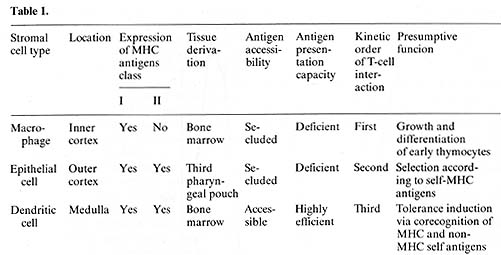
            Intrathymic antigen presentation was assayed by coculture of antigen-specific 
              l-Arestricted cloned T -helper cells with purified irradiated thymic 
              Iymphostromal-cell complexes [II, 15]. As antigens we used myoglobin, 
              L-glutamic acid high 60-L-alanine high 30 L-tyrosine high 10 (GAT), 
              and keyhole limpet hemocyanin (KLH). Proliferation of T cells was 
              measured 72 h after culture in vitro by (³H]thymidine uptake. Antigen 
              was either injected intravascularly (i.v.) before isolation of the 
              stimulator population or added to the culture in vitro. After injection 
              of 0.5 mg myoglobin/g body weight i.v. in to C57BL/Ka mice, T-ROS 
              copurified with specific stimulation of l-A b-restricted myo globin-specific 
              T-helper cells. This antigenspecific stimulation was a property 
              of Thy1.2-negative stromal cells (anti- Thy 1.2 antibody plus complement 
              treatment did not alter the presentation capacity of T-ROS) and 
              could be inhibited by more than 90% after pretreatment of the stimulator 
              population with anti l-Ab monoclonal antibody and complement. Antigen 
              traffic to the thymus in vivo was dose dependent within the range 
              of 1.0-0.25 mg m yoglobin/g body weight. Threshold doses for thymic 
              and splenic antigen-presenting cells (A PC) required to present 
              antigen were similar, indicating no significant seclusion in vivo 
              of A PC enclosed in the T-ROS fraction. Similar results were obtained 
              after injection of KLH (molecular weight 3 x10 high 6) and GAT (molecular 
              weight, I x10 high 5) [ 15]. When kinetics of antigen persistence 
              in the thymus were measured, antigen-specific stimulation of T cells 
              was demonstrable up to 48 h after injection i.v. (Fig. IA). The 
              prolonged presence of antigen within the thymus argues against a 
              trivial explanation of these resuIts, namely the uptake of antigen 
              by stromal cells (which were secluded in the intact organ), after 
              disruption of the tissue context during the isolation procedure. 
              Although myoglobin (molecular weight 17000) is rapidly cleared from 
              the circulation, the APC activity of T -ROS was unchanged when tested 
              15 min or 12 h after injection of antigen.  


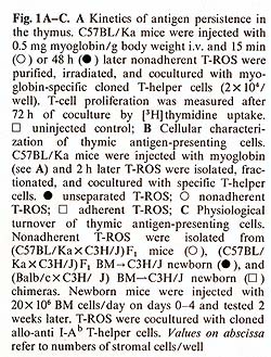
            In further defining the cell type(s) responsible for uptake of antigen 
            in vivo and presentation in vitro we separated T-ROS stromal cells 
            into adherent and nonadherent fractions. More than 90% of adherent 
            stromal cells are composed of I-A/ E-negative, F 4/80-positive, phagocytic 
            MØlike cells, whereas the nonadherent stromal cells contain 
            50%-80% nonphagocy tic F 4/80-negative, I-A/E-positive DClike cells 
            [I, 14]. When separately tested for APC activity after injection of 
            myoglobin i.v., the nonadherent fraction contained all functional 
            APC, whereas the adherent-cell fraction even when pulsed with additional 
            antigen in vitro remained nonstimulatory (Fig. 1 B). Though this separation 
            method needs further confirmation by enrichment protocols using strictly 
            lineage-specific surface markers. it was reproducibly found that depletion 
            of strongly adherent MØ did not affect the ability of the T 
            -ROS fraction to present antigen. The lack of class I I MHC-antigen 
            expression on cortical macrophages forming T -ROS is compatible with 
            this result. In order to test DCs and TNCs separately for their accessibility 
            and capacity to present antigen, we turned our attention to their 
            different embryonic origins. DCs are strictly bone marrow derived 
            whereas epithelial cells are derived from the third pharyngeal pouch. 
            Thus, PI> (P1 X P2) F1 radiation chimeras were analyzed in which DCs 
            were com pletely replaced by P1-type cells and epithelial cells remained 
            of the F1 type. When antigen presentation by cells isolated from such 
            animals was tested 10 weeks after reconstitution with T -helper cells 
            restricted to P2type I-A antigens, no T -cell proliferation was measured 
            using either purified T -ROS (that is bone-marrow-derived stromal 
            cells) or epithelial cells as stimulators. The latter, however, expressed 
            class II-MHC-determinants of P2-type abundantly, as determined by 
            fluorescence microscopy. This lack of antigen presentation by thymic 
            epithelial cells after injection of myoglobin i.v. could not be overcome 
            by providing optimal doses of antigen in vitro [15]. This result indicates 
            an intrinsic deficiency of epithelial cells in stimulation of T-helper 
            cells, rather than a seclusion from antigen in vivo. It is not clear 
            to date whether the epithelial cells lack the ability to process antigen 
            and/ or fail to produce obligatory costimulation factors (e.g., interleukin-I). 
            Interestingly, thymic epithelial cells have been successfully grafted 
            across allogeneic barriers without being rejected [21]. Given the 
            assumption that DCs are responsible for antigen presentation in the 
            thymus, we further assessed the physiological turnover of thymic DCs 
            (equivalent here to nonadherent T-ROS). To this end we used nonradiation 
            chimeras. Newborn P1 mice were given multiple injections with F1 bone 
            marrow cells at daily intervals. Such animals establish a stable bone 
            marrow chimerism which is proportional to the dose of donor cells 
            injected (Kyewski, unpublished data). In such "normal" hosts, without 
            prior ablation of bone marrow-derived hemopoietic lineages, F1-BM-derived 
            cells establish stem-cell chimerism and replace host cells during 
            physiological turnover. When nonadherent T-ROS from such F1> P1 newborn 
            chimeras were cocultured with cloned T -helper cells restricted to 
            P2-type I-A antigens, specific proliferation was detected (Fig. I 
            C). This proliferative response amounted to about 10%-20% of the magnitude 
            induced by normal F1 mice-derived T -ROS, indicating a significant 
            replacement in the thymus of host-type DCs by cells of donor origin. 
            The result indicates that medullary DCs, in contrast to cortical epithelial 
            cells, undergo a constant physiological turnover and replacement by 
            extrathymic DCs. Thus, in addition to the direct entry of blood-bome 
            antigens into the thymus, circulating antigen-Iaden-DCs may contribute 
            to the spectrum of intrathymically presented antigens. The described 
            results, in concert with earlier studies on these cell interactions 
            [25, 13-16, 7], indicate a strict compartmentalization of thymic stromal 
            cells with regard to their accessibility to circulating antigens and 
            their intrinsic capacity to present these antigens to T cells. Macrophages 
            and epithelial cells (here isolated by virtue of their interactions 
            with thymocytes in vivo) seem to be highly inefficient in presentation 
            of soluble protein antigens and are presumably secluded from blood-borne 
            antigens by avascular blood-cortex barrier. In contrast, presenting 
            DCs are strictly confined to the medulla, which in turn displays a 
            vascular architecture permissive to the passage of macromolecules 
            [20]. Thus, an important aspect of cortex/medulla dichotomy with regard 
            to T -cell recognition resides in either the prevention or facilitation 
            of T-cell encounters with non-MHC antigens in conjunction with self 
            MHC-antigens. In the following we speculate on the possible roles 
            of the three recognition steps in the context of the development of 
            the T -cell repertoire. Pre- T cells probably enter the thymus at 
            the cortical side of the corticalmedullary junction and first interact 
            with and proliferate around macrophages in the inner cortex (Fig.2). 


              The role of these macrophages may resemble those in hemopoietie 
              islands. in the bone marrow, in which they support the growth and 
              differentiation of erythropoietic and granulopoietic cell lineages 
              by direct cell-cell contact [2, 5]. This early stage of thynlocyte-accessory-cell 
              interaction may not yet involve the T -cell receptor for antigen 
              recognition but may induce its expression. Affer this initial MØ-induced 
              growth and differentiation phase a second interaction with epithelial 
              cells follows in the outer cortex. As proposed previously. this 
              in teraction ma y represent the recognition of self-MHC determinants 
              expressed on epithelial cells by a minor selective set of clonally 
              expressed T-cell receptors, thus determining the selfMHC restriction 
              of cytotoxic and helper T cells [6, 26]. If correct, one would predict 
              that all T cells associated with epithelia] cells should have productively 
              rearranged T-cell-receptor genes and express membrane-bound T-cell-receptors 
              [ 12]. In the absence of non-MHC antigens this recognition step 
              has to be different from the obligatory corecognition of MHC antigens 
              and nominal antigens exhibited by mature T cells [23, 3]. A third 
              type of intercellular recognition, now in the medulla between thymocytes 
              and dendritic cells, displays the characteristic MHC-restricted 
              recognition of non-MHC antigens, which enter this compartment and 
              are presented here to maturing T cells. By conjecture this site 
              may constitute a possible microenvironment where self-tolerance 
              is induced. This proposition receives indirect support fronl the 
              recent observation that thyn1ic epithelial cells when grafted across 
              allogeneic barriers do not induce T-cell tolerance to their own 
              class II MHC antigens [21]. DCthymocyte interactions would conform 
              to the prediction that tolerance induction is MHC restricted [8, 
              18, 19]. In addition, the ontogeny of thymocyte-DC interactions 
              parallels the induction of self-tolerance [ 17] (Kyewski, unpublished 
              data). These considerations, however, leave the cellular and molecular 
              mechanisms of how self-tolerance is induced and maintained completely 
              unexplained. According to this model, recognition structures on 
              stromal cells select for complementary receptors on T cells expressed 
              at the respective stage of their interaction. With the advent of 
              monoclonal antibodies and molecular probes specific for the T -cell-receptor 
              certain predictions of this model nlay be tested [9, 10]. In the 
              absence of direct evidence for a precursor-product relationship 
              between the various interactions, and the unknown fate of the selected 
              thymocytes, the developmental pathways of T cells still remain conjectural 
              [22] ( Fig. 2). The outlined results and speculations depict the 
              com plexity of intra thymic l-cell maturation with regard to cell-to-cell 
              communication, compartmentalization, and directed lymphocyte traffic. 
              The possibly critical involvement of stromal cells in growth control 
              of hematopoietic cell lineages has recently been reemphasized [4]. 
              In view of the notion that some forms of acute T-cellleukemias may 
              represent an arrest in differentiation rather than an irreversible 
              transformation step, the analysis of the inductive signals responsible 
              for T -cell growth and differentiation may aid our understanding 
              of the mechanism of T -cell leukemogenesis. T -cell transformation, 
              at least in the murine model, has been shown to be strictly dependent 
              on an intact thymic microenvironmen t.  
             
              Acknowledgment.  
               
              This work has been conducted under the stimulating guidance of the 
              late Henry S. Kaplan, Cancer Biology Research Laboratory, Dept. 
              of Radiology in collaboration with Dr. C. G. Fathman, Div. of Immunology, 
              Dept. of Medicine, Stanford University Medical School. The author 
              was supported by a postdoctoral fellowship of the DFG. 
             
              References  
            1. Austyn JM, Gordon S (1981) F4/80, a mono clonal antibody directed 
              specifically against the mouse macrophage. Eur J Immunol 10: 805-811 
              2. Een-Ishay Z, Yoffey JM ( 1972) Ultrastructural studies of erythroblastic 
              islands of rat bone marrow. II Resumption of erythropoiesis in erythropoietically 
              depressed rebound marrow. Lab Invest 26:637-647 
              3. Eevan MJ (1981) Thymic education. Immunol Today 2: 216-219  
              4. Eoettiger D, Anderson S, Dexter TM ( 1984) Effect of ssrc infection 
              on long-term marrow culture: increased self renewal of hemopoietic 
              progenitor cells without leukemia. Cell 36:763-773  
              5. Cline MJ, Golde DW (1979) Cellular interactions in hematopoiesis. 
              Nature 277: 177-181  
              6. Fink PJ, Bevan MJ (1978) H-2 antigen of the thymus determines 
              lymphocyte specificity. J Exp Med 148: 766- 775  
              7. Fink PJ, Weissman IL, Kaplan HS, Kyewski EA (1984) The immunocompetence 
              of murine stromal cell-associated thymocytes. J ImmunoI132:2266-2272 
               
              8. Groves ES, Singer A (1983) Role of H-2 complex in the induction 
              of T cell tolerance to minor histocompatibility antigens. J Exp 
              Med 158:1483-1497  
              9. Haskins K et al. (1983) The major-histocompatibility-restricted 
              antigen receptor on T cells. I. Isolation with a monoclonal antibody.JExp 
              Med 157:1149-1169  
              10. Hedrick Set al. (1984) Isolation of cDNA clones encoding T -cell-specific 
              membraneassociated proteins. Nature 308: 149-153  
              11. Infante AJ, Atassi MZ, Fathman CG (1981) T cell clones reactive 
              with sperm whale myoglobin. Isolation of clones with specificity 
              for individual determinants on myoglobin. J Exp Med 154:1342-1356 
               
              12. Kavaler J, Davis MM, Chien Y -H (1984) Localization of a T -cell 
              receptor diversity-region element. Nature 310:421-423  
              13. Kyewski BA, Kaplan HS (1982) Lymphoepithelial interactions in 
              the murine thymus: phenotypic and kinetic studies on thymic nurse 
              cells. J ImmunoI128:2287-2294  
              14. Kyewski BA, Rouse RV, Kaplan HS (1982) Thymocyte rosettes: multicellular 
              complexes of lymphocytes and bone-marrow-derived stromal cells in 
              the murine thymus. Proc Natl Acad Sci USA 79: 5646-5650  
              15. K yew ski EA, Fathman CG, Kaplan HS ( 1984 a) Intrathymic presentation 
              of circulating non-major histocompatibility complex antigens. Nature 
              308: 196-199  
              16. Kyewski BA, Travis M, Kaplan HS (1984b) Intrathymic lymphopoiesis: 
              stromal cell-associated proliferation of T cells is independent 
              of lymphocyte genotype. J Im munol133: 1111-1116  
              17. Lu C-Y, Eeller Dl, Unanaue E (1981) During ontogeny, Ia-bearing 
              accessor cells are found early in the thymus but late in the spleen. 
              Proc Natl Acad Sci USA 77: 1597 -1601  
              18. Matzinger P, Zamoyska R, Waldmann H (1984) Self tolerance is 
              H-2 restricted. Nature 308:738-741 
              19. Rammensee, H-G, Eevan MJ (1984) Evidence from in vitro studies 
              that tolerance for self-antigens is MHC-restricted. Nature 308: 
              741-744  
              20. Raviola E, Karnovsky MJ (1972) Evidence for a blood-thymus barrier 
              using electron opaque tracers. J Exp Med 136:466-498 
              21. Ready AR, Jenkinson EJ, Kingston R, Owen JJT (1984) Successful 
              transplantation across major histocompatibility barrier of deoxyguanosine-treated 
              embryonic thymus expressing class II antigens. Nature 310: 231-233 
               
              22. Scollay R (1983) Intrathymic events in the differentiation of 
              T lymphocytes: a continuing enigma. Immunol Today 4:282-286 
              23. Yon Böhmer H, Haas W, Jerne NK (1978) Major histocompatibility 
              complex-linked immune-responsiveness is acquired by lymphocytes 
              of low-responder mice differentiating in thymus of high-responder 
              mice. Proc Natl Acad Sci USA 75: 2439-2442  
              24. Wagner H, Röllinghoff M, Pfitzenmaier K (1982) The influence 
              of the thymus on the generation of T cell repertoire. Behring Inst 
              Mitt 70  
              25. Wekerle H, Ketelsen U-P, Ernst M (1980) Thymic nurse cells. 
              Lymphoepithelial cell complexes in murine thymus: morphological 
              and serological characterization. J Exp Med 151:925-944  
              26. Zinkernagel RM et al. (1978) On the thymus in the differentiation 
              of "H-2 self recognition " by T cells: evidence for dual recognition. 
              J Exp Med 147: 882-896  
           |